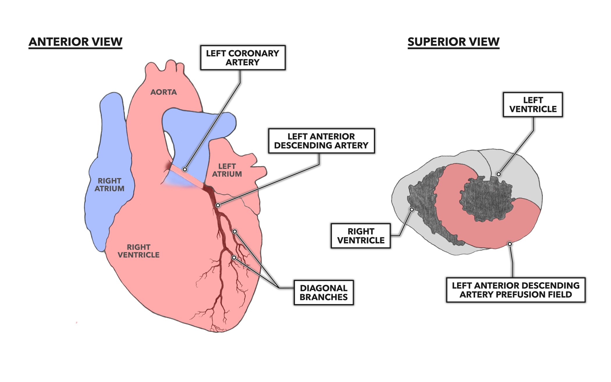
Number Of Diagonal Arteries / Crossfit The Heart Part 7 Coronary Circulation. There are multiple smaller coronary arteries that branch off from the left and right coronary artery and even the circumflex artery. If a ramus intermedius artery is present, the diagonal arteries are. In about 80% of people, the lad wraps around the bottom of the heart and supplies the area beyond that. The main coronary arteries are: Lv function, including ejection fraction and wall motion, should also be assessed.
Coronary circulation is the circulation of blood in the blood vessels that supply the heart muscle (myocardium). Typically, the number of diagonal and marginal branches is specified and their disease burden quantified. Blockage of the lad artery significant blockages of the lad artery can be dangerous simply because the lad supplies such a large territory. Time, and the number of devices. The main coronary arteries are:
Crossfit The Heart Part 7 Coronary Circulation from www.crossfit.com The right coronary artery divides into smaller branches, including the right posterior descending artery and the acute marginal artery. 26 the septal arteries are the basis of efficient intercoronary collateralization between the lad and pda and probably the most common type. This cohort study investigated 449 consecutive chinese, 250 cases with cad and 199 without cad, who were certified by coronary artery angiography in our center. The number of intersection points made by the diagonals of a regular polygon in 1997. The coronary arteries wrap around the outside of the heart. The number of superior septal arteries ranges from 1 to 10, with an average of 4.2, and the inferior one's ranges from 0 to 7, with an average of 2. The diagonals initially run on the surface of the heart and therefore can be visualized by ct imaging. Coronary arteries supply oxygenated blood to the heart muscle, and cardiac veins drain away the blood once it has been deoxygenated.
The number of intersection points made by the diagonals of a regular polygon in 1997.
They often run parallel to one another and are variable in number (often 2 to 9). The new guideline applies to the number of coronary arteries treated and not the number of sites treated. There are multiple smaller coronary arteries that branch off from the left and right coronary artery and even the circumflex artery. This third branch originates between the angle formed by the lad and the left circumflex arteries and has various names, including ramus intermediate, median artery, left diagonal artery, and straight left ventricular artery. left anterior descending artery. A reporting system on patients evaluated for coronary artery disease. In general, the diagonal branches are larger, longer, and have more angular takeoff from the lad artery. Over 99% of people have at least one diagonal branch of the left anterior descending artery. Characteristic differences and the relation of delc to cad were assessed by. Time, and the number of devices. The myocardium itself actually gets 100% of its oxygen and nutrient supply from the coronary arteries and receives no oxygen or nutrients from the blood that passes through the ventricls of the heart. The diagonal branches had a diameter greater than 1.5 mm. This allows blood to flow around the blocked artery to another artery nearby. If a ramus intermedius artery is present, the diagonal arteries are.
Blockage of the lad artery significant blockages of the lad artery can be dangerous simply because the lad supplies such a large territory. Most frequently there is atleast one diagonal branch. Left coronary artery left circumflex. Many reports have claimed associations between diagonal earlobe crease (delc) and coronary artery disease (cad), but data in chinese populations are limited. Failure to visualize any diagonal artery denotes occlusion.
Is Septal Branches Of Left Anterior Descending Coronary Artery Immune To Atherosclerosis Dr S Venkatesan Md from drsvenkatesan.files.wordpress.com Each number is the numbers directly. The total number of elements of the given matrix will not exceed 10,000. When the coronary arteries narrow to the point that blood flow to the heart muscle is limited (coronary artery disease), collateral vessels may enlarge and become active. The first diagonal (d1) branch tends to be the most prominent. Narrowing of the arteries can be caused by a process known as atherosclerosis (most common), arteriosclerosis, or arteriolosclerosis.this occurs when plaques (made up of deposits of cholesterol and other substances) build up over time in the walls of the arteries. Collateral circulation is a network of tiny blood vessels, and, under normal conditions, not open. Because the rest of the body, and most especially the brain, needs a steady supply of oxygenated blood that is free of all but the slightest. If a ramus intermedius artery is present, the diagonal arteries are less prominent and arise more distally.
Each artery is a muscular tube lined by smooth tissue and has three layers:
Each number is the numbers directly. Each artery is a muscular tube lined by smooth tissue and has three layers: Collateral circulation is a network of tiny blood vessels, and, under normal conditions, not open. When the coronary arteries narrow to the point that blood flow to the heart muscle is limited (coronary artery disease), collateral vessels may enlarge and become active. Blockage of the lad artery significant blockages of the lad artery can be dangerous simply because the lad supplies such a large territory. Then codominent vessel proximal right coronary artery has 90% disease. The main coronary arteries are: The second of the three longest branches off of the left anterior descending artery which supplies the anterolateral wall of the left ventricle. If a ramus intermedius artery is present, the diagonal arteries are less prominent and arise more distally. A reporting system on patients evaluated for coronary artery disease. Lv function, including ejection fraction and wall motion, should also be assessed. This allows blood to flow around the blocked artery to another artery nearby. Coronary circulation is the circulation of blood in the blood vessels that supply the heart muscle (myocardium).
8 a lesion was considered hemodynamically significant when its diameter was reduced by at least 65%. Characteristic differences and the relation of delc to cad were assessed by. The diagonals initially run on the surface of the heart and therefore can be visualized by ct imaging. Then codominent vessel proximal right coronary artery has 90% disease. Many reports have claimed associations between diagonal earlobe crease (delc) and coronary artery disease (cad), but data in chinese populations are limited.
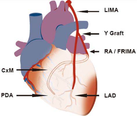
Why And How To Achieve Total Arterial Revascularisation In Coronary Surgery from oaepublishstorage.blob.core.windows.net Coronary artery disease (cad) is a major cause of mortality, resulting in an estimated 7.6 million deaths every year all over the world. 1 conventional coronary angiography (cca) is the only undisputed means of visualizing the coronary arterial system in vivo and is regarded the gold standard for the assessment of modality of coronary arteries since cca was first discovered by. In an rao projection, this artery often arises where the left anterior descending angles toward the apex. Смотреть что такое diagonal arteries в других словарях: Each artery is a muscular tube lined by smooth tissue and has three layers: Failure to visualize any diagonal artery denotes occlusion. Many reports have claimed associations between diagonal earlobe crease (delc) and coronary artery disease (cad), but data in chinese populations are limited. The diagonal branches had a diameter greater than 1.5 mm.
If a ramus intermedius artery is present, the diagonal arteries are less prominent and arise more distally.
They had 40 lad/diagonal branch bifurcation lesions. This allows blood to flow around the blocked artery to another artery nearby. The main coronary arteries are: Blockage of the lad artery significant blockages of the lad artery can be dangerous simply because the lad supplies such a large territory. Additional smaller branches of the coronary arteries include the obtuse marginal (om), septal perforator (sp), and diagonals. (ii) only the morphologically left ventricle receives the diagonal arteries from the anterior and posterior interventricular arteries. Typically, the number of diagonal and marginal branches is specified and their disease burden quantified. Each number is the numbers. Then codominent vessel proximal right coronary artery has 90% disease. Each number is the numbers directly. In about 80% of people, the lad wraps around the bottom of the heart and supplies the area beyond that. Each artery is a muscular tube lined by smooth tissue and has three layers: The second of the three longest branches off of the left anterior descending artery which supplies the anterolateral wall of the left ventricle.
Share :
Post a Comment
for "Number Of Diagonal Arteries / Crossfit The Heart Part 7 Coronary Circulation"


Post a Comment for "Number Of Diagonal Arteries / Crossfit The Heart Part 7 Coronary Circulation"